MMI announces global imaging services collaboration with Micron
Learn MoreThe MMI Advantage
Experts in Abdominal and
Body Imaging
Our reviewers are experts in thoracic, abdominal and pelvic imaging including but not limited to liver, pancreas, prostate, kidney, bladder, breast imaging.
Sophisticated and Validated Image Analysis Solutions
We conduct all of our analysis using sophisticated tools, workflows and methodologies that are specifically validated for clinical trial needs.
Customized Analysis Protocols for Your Trial Needs
Our team of scientists work closely with you to provide guidance and are flexible to customize your analysis requirements.
Experience and Expertise
Modality Expertise
Ultrasound
X-ray
CT
CTA
MRI
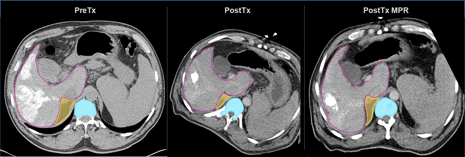
MMI has over twenty years of experience in providing core lab support for more than 150 device and biopharmaceutical companies internationally. We have developed custom methodologies in support of multiple regulated clinical trials using validated third-party tools that have been integrated into MMI’s proprietary system for advanced image analysis and 3D volumetric quantification.
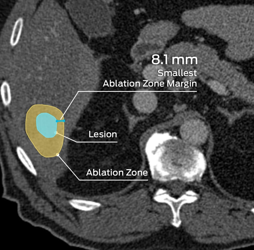
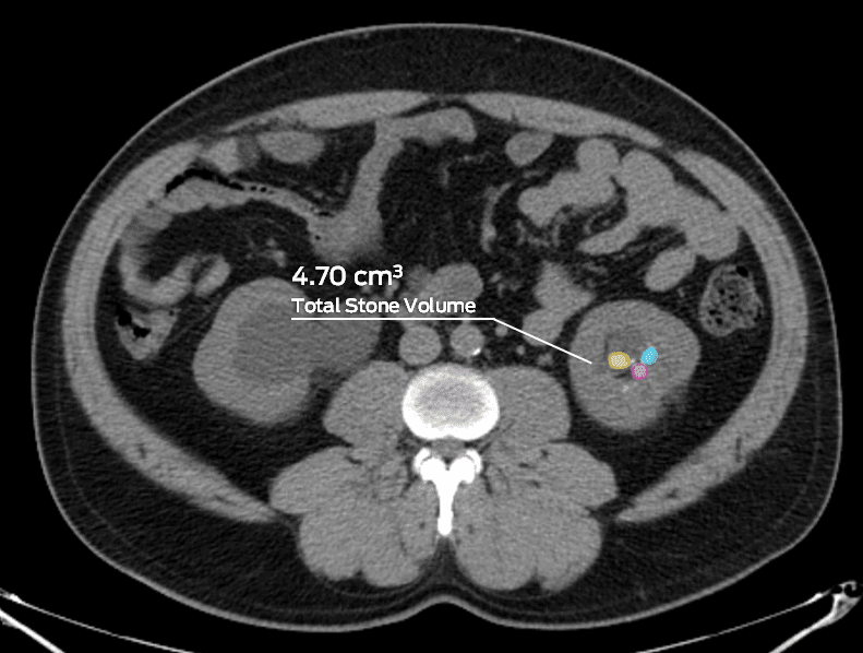
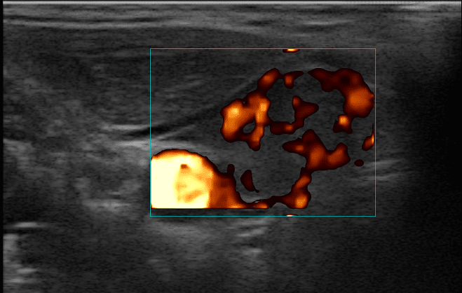
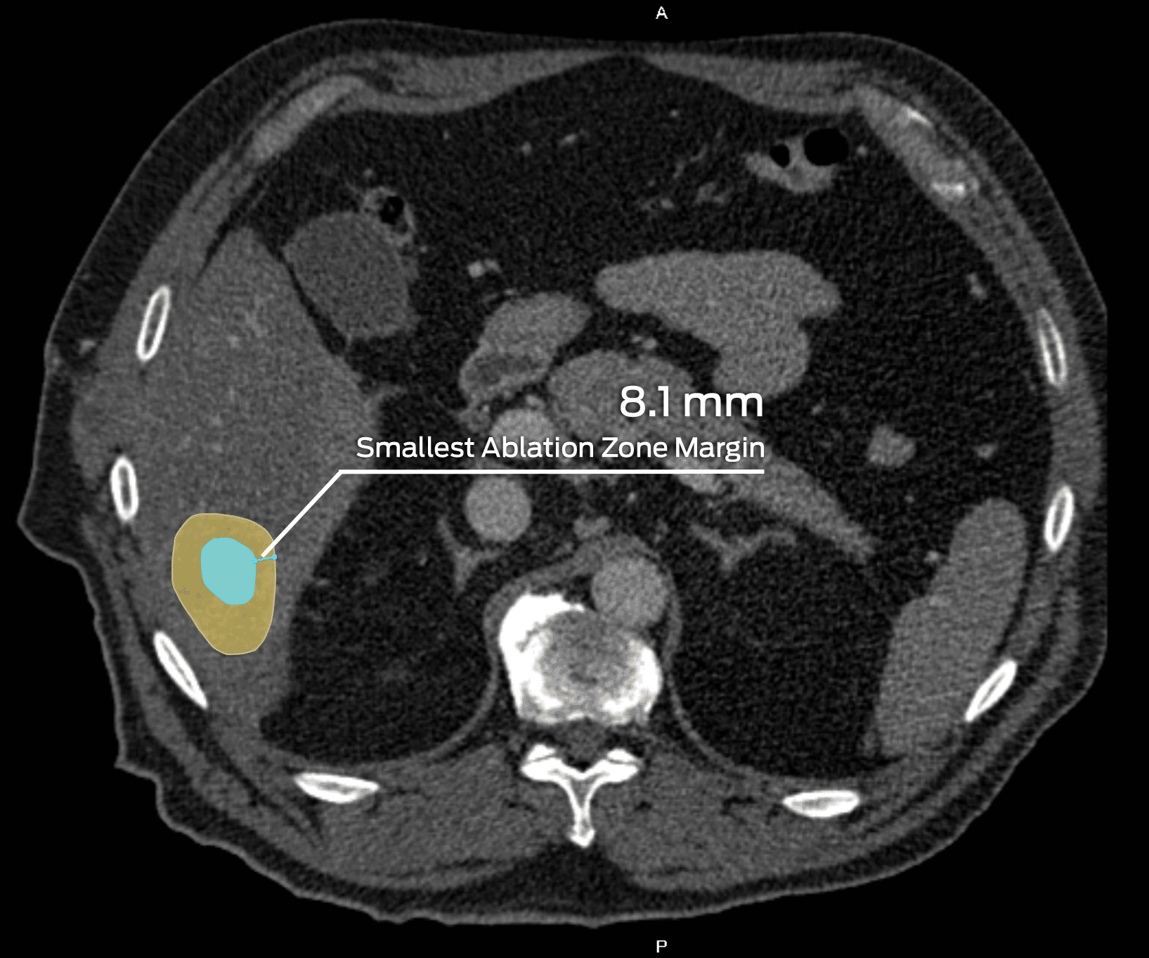
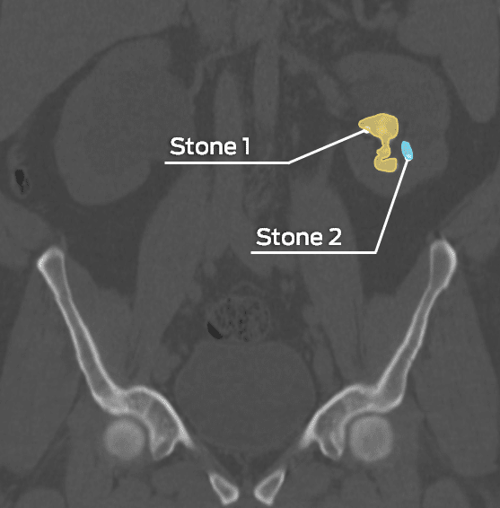
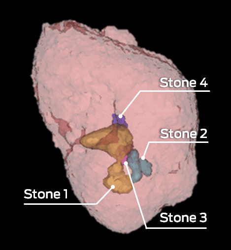
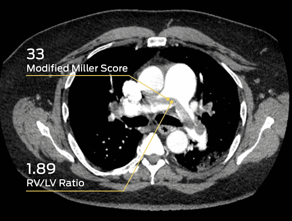
Our team of clinical and scientific experts in abdominal & pelvic imaging and medicine includes editors of high-impact journals, department chairs of radiology, and division chiefs of surgery. Our depth and diversity of experts allow us to support virtually all of the major imaging modalities in one core lab. MMI’s experience in thoracoabdominal studies include: lung, breast and thyroid nodule detection and evaluation, liver and lung tumor ablation, kidney and urinary stone removal, colorectal anastomoses, hemorrhage detection systems and vascular embolization devices.
WHAT WE OFFER
Analysis Capabilities
Some of Our Experts
Anup Shetty, MD
Associate Professor of Radiology, Fellowship Director for Advanced Abdominal Imaging, Service Director for Body MRI, Mallinckrodt Institute of Radiology, Washington University School of Medicine
Following an internal medicine residency at the Washington University School of Medicine, Dr. Shetty completed a diagnostic radiology residency and body magnetic resonance imaging fellowship at Mallinckrodt Institute of Radiology (MIR). His areas of clinical and research interest include abdominal imaging, body MRI, emergency radiology, vascular imaging, genitourinary imaging, prostate MRI and radiology education. In 2019, Dr. Shetty was named an inaugural fellow of the School of Medicine’s Academy of Educators, and received the 2021 Rising Star Award. In 2022, he was named the Teacher of the Year for the MIR and an Honored Educator by the Radiological Society of North America (RSNA). He also serves on several committees, including as Chair of the American Board of Radiology Online Longitudinal Assessment GI Committee. Dr. Shetty has also published numerous research and educational works in the field of body imaging over the past decade.
Atif Zaheer, MD
Director of Cross-Sectional Body Imaging Fellowship Program, Department of Radiology and Radiological Science, Johns Hopkins University
Dr. Zaheer is board-certified in diagnostic radiology and practices at the Johns Hopkins Hospital in Baltimore, MD. He earned his medical degree from the Aga Khan University and completed a residency in diagnostic radiology at Beth Israel Deaconess Medical Center/Harvard Medical School, followed by a fellowship in abdominal imaging and interventional radiology at Brigham and Women’s Hospital/Harvard Medical School. Dr. Zaheer is clinically active in cross-sectional imaging of the body using CT, MRI and ultrasound and is also involved in multidisciplinary conferences for pancreas, liver and rectal cancer imaging. His research interests include imaging of tumors and inflammatory disorders of the pancreas and is considered a leader in pancreatic imaging. He is the Associate Editor of the Journal of Abdominal Radiology and actively participates in clinical trials, publishes scientific articles, authors books on pancreatic and gastrointestinal imaging, and gives talks at national and international conferences.
Relevant Scientific Resources
Key Publications Involving MMI
Let MMI provide insights into your clinical study imaging.
Have questions? We’ll connect you immediately to one of our scientific managers and imaging experts. Your time is precious, and we want to make the most out of it.
