MMI announces global imaging services collaboration with Micron

Learn More

Spine

Spine
Spine
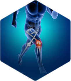
Orthopedics
Orthopedics
Cardiovascular
Cardiovascular
Otorhino – laryngology
Neurology
Neurology
Thoraco – abdominal
Thoraco – abdominal
Spine
Spine
Group 2096

The MMI Advantage

Spine 1
Leading spine
imaging core lab

We estimate MMI has been involved in ~80% of regulated spine trials run in the U.S. over the past 20 years.

Spine 2
Gold standard QMA® image analysis technology

Our patented QMA® technology has the highest accuracy and reproducibility of any published method for quantitative spine measurements and has been referenced in over 200 publications.

Spine 3
Experienced with all types of spine therapies

Our staff and expert reviewers have extensive experience across the full spectrum of spine treatments, including fusion, motion preservation, augmentation, and deformity correction.

Experience and Expertise
MMI’s scientific experts have contributed hundreds of publications to the scientific literature on spine imaging and research. No other core lab offers our unique combination of experienced readers, scientific expertise, and specialized technology for spine clinical trials.
TA_Spine

Modality Expertise

X-ray
MRI
CT
Ultrasound
MMI has extensive experience with motion preserving technology, including total disc replacement, facet replacement, disc repair and regeneration, and dynamic stabilization therapies. Our spine fusion experience includes assessments of allografts, autografts, and bone graft substitutes. We also have experience with deformity correction, interspinous distraction, and fracture stabilization and healing technologies. We are leaders in the evaluation of biologic products for bone and soft-tissue repair and have evaluated an array of novel treatments to slow or reverse disc degeneration and promote bone healing following spine fusion procedures.

We are experienced in virtually all major quantitative and semi-quantitative scoring systems to assess disease severity and response to therapy from medical imaging. We support image calibration strategies through the use of XCalibR, MMI’s proprietary X-ray Calibration Ring marker. When appropriate, we apply validated, computer-assisted methods to objectively document the effect of new treatments. MMI’s proprietary and patented QMA® technology produces accurate and reproducible measurements from spine radiographic images and has been published in over 200 peer-reviewed articles. Learn more about QMA®.

WHAT WE OFFER

Analysis Capabilities

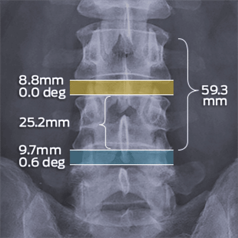
Quantitative spine measurements (e.g. rotation, translation, center of rotation, disc height, lordosis)
Spinal stability and alignment, including deformity correction (e.g. sagittal balance, Cobb angle)
Bony bridging and fusion, including extent of bony integration
Bone density and volume from QCT

Quantification of bone formation or removal
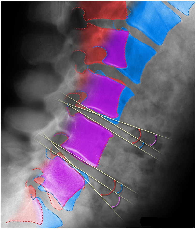
Structural bony anatomy (e.g. bone defects, osteophytes, Schmorl’s nodes, stenosis)
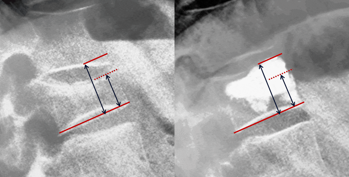
Vertebral augmentation and height restoration
Fracture and fracture healing
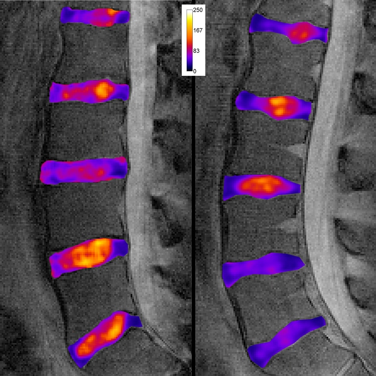
Disc composition and hydration, including qualitative, semi-quantitative, and relaxation mapping techniques (e.g. T2 mapping, T1ρ mapping)
Soft tissue structure and morphology (including annulus, nucleus, ligaments)
Position and volume changes of injectable materials
Implant stability and fixation
Implant interface remodeling
Custom implant analyses (e.g. preoperative planning, custom assessment development)
High-precision measurements of disc angle in flexion and extension using MMI’s proprietary QMA® analysis technology
High-precision measurements of disc angle in flexion and extension using MMI’s proprietary QMA® analysis technology
MR relaxation mapping to quantify disc quality during repair and regeneration
3D render of spine fusion to evaluate device and hardware positioning, bone-implant interface changes, implant integrity, and more
Measurement of vertebral body height restoration after vertebroplasty
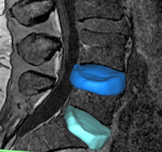
Segmented disc volume overlaid on MR image slice
Quantity and quality of spine motion (e.g. angular motion, center of rotation) following total disc replacement, evaluated using MMI’s proprietary QMA® analysis technology
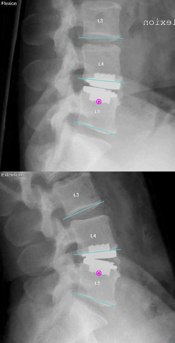
Quantity and quality of spine motion (e.g. angular motion, center of rotation) following total disc replacement, evaluated using MMI’s proprietary QMA® analysis technology
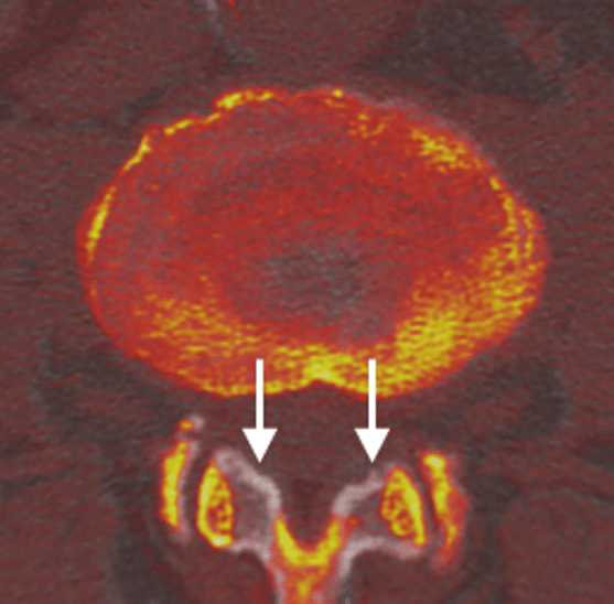
PostOp (color) CT image overlaid on the PreOp (grayscale) image, showing regions of bone that were removed during decompression (white arrows).
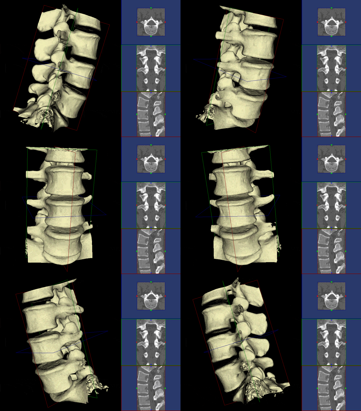
3D CT visualization to assess form and function of the spine, including changes due to treatment

Some of Our Experts

John Hipp, PhD

Co-founder

Dr. Hipp is the Former Director of the Spine Research Lab at Baylor College of Medicine. He has over 30 years’ experience in quantitative imaging methods for spine research and is a recognized leader in spine related research. He has published over 100 imaging-related papers and book chapters, managed development of more than 75 spine-related imaging charters, and is a reviewer for eight leading journals including Spine and The Spine Journal. For his outstanding contributions, Dr. Hipp has received the prestigious AAOS Kappa Delta Orthopedic Research Award and NASS 2022 Henry Farfan Award.

Mark Schweitzer, MD

Vice President of Health Affairs,
Wayne State University

Dr. Schweitzer is fellowship-trained in osteoradiology, and has over 30 years’ experience in research, education, and clinical practice. Dr. Schweitzer is the former presiding officer of the Radiological Society of North America (RSNA) and International Skeletal Society (ISS), is the current Editor-in-Chief of JMRI, and serves on the Editorial Board of MRM. He has published over 400 peer-reviewed papers, 68 book chapters and 4 books, and delivered 800 lectures in 38 countries. Dr. Schweitzer is also an experienced scientific advisory panel member for FDA and NIH.

Documentation

MMI Bibliography of Spine Imaging Studies
Lorem ipsum dolor sit amet consectetur.
Spine CAMP™ Instructions for Use

Let MMI provide insights into your clinical study imaging.

Have questions? We’ll connect you immediately to one of our scientific managers and imaging experts. Your time is precious, and we want to make the most out of it.
Contact icon