MMI announces global imaging services collaboration with Micron

Learn More

Ear, Nose, & Throat

Spine
Spine
Orthopedics
Orthopedics
Cardiovascular
Cardiovascular
Otorhino – laryngology
ENT
Neurology
Neurology
Thoraco – abdominal
Gastrointestinal & Urology
Group 2096

The MMI Advantage

Group 2049
State-of-the-art volumetric analysis capability

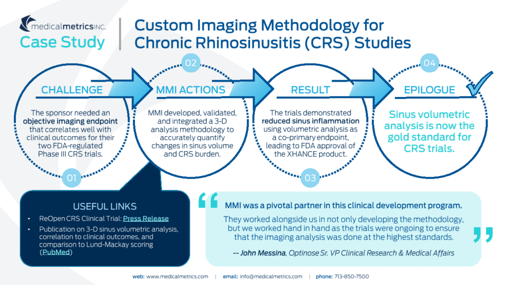
Through collaborations with industry experts and key opinion leaders, MMI has developed validated, custom methodologies to precisely quantify the mucosal thickening or inflammatory disease associated with Chronic Rhinosinusitis (CRS).

ENT 1
Experienced with multiple CRS trials

We are a preferred imaging core lab for CRS trials and have the ability to objectively characterize sinus opacification to detect minute changes that can be missed using traditional qualitative grading systems. Furthmore, MMI has supported trials involving wound healing and nasal polyp characterization.

ENT 3
Excellent industry
reputation

“We are blessed to have a partnership with such a great leader in CT assessment within the ENT community.  The novelty and the quality of work done by Medical Metrics is unprecedented, which is instrumental for us to generate a high impact publication and awareness from ENT physicians.”  - Clinical Research Manager from Top 5 Medical Device Companies

Experience and Expertise

MMI is a leading core lab for ENT trials and has developed an objective measure for characterizing sinus opacification as an imaging endpoint. 

TA_ENT

Modality Expertise

CT
MRI
Endoscopy

MMI has worked with leading pharmaceutical and device companies in the ENT space to study treatment effects in a broad subject population that includes CRS, CRS + polyposis, FESS, and ethmoidectomy. Our experience includes evaluation of drug-eluting stents, irrigation cannulas administered through the lacrimal duct, drug exhalation systems, and numerous biologics.

We have authored a variety of radiographic protocols, guidance materials, and justification documents to validate the methodologies used to quantify mucosal thickening, including the use of volumetric analysis in regulated clinical trials. MMI also has the ability to provide quick-turn analysis in support of subject eligibility screening and has experience conducting analyses in thousands of subjects in support of regulated clinical trials.

WHAT WE OFFER

Analysis Capabilities

CRS (e.g. Lund-Mackay, Zinreich, OMC patency)

Nasal polyp characterization (e.g. NPS & other variants)

Assessment of wound healing (e.g. Olfactory Fossa depth)

Sinus anatomy characterization (e.g. depth of the Olfactory Fossa)
Volumetric quantification of mucosal thickening (maxillary, ethmoid, nasal cavity, frontal sinus ostium, middle meatus, OMU)

Mucosal modeling to evaluate disease burden and progression over time

Modeling of airway restrictions and central canal stenosis

3D modeling of the maxillary and ethmoid sinuses

Zinreich scores of right anterior and posterior ethmoid sinuses on sagittal CT

Zinreich scores of right anterior and posterior ethmoid sinuses on sagittal CT

Digital subtraction technique for determining the difference between baseline and post-treatment images to identify areas of reduced sinus opacification

Digital subtraction technique for determining the difference between baseline and post-treatment images to identify areas of reduced sinus opacification

Quantification of ethmoid (yellow) and maxillary (blue) sinus opacification in coronal CT

Quantification of ethmoid (yellow) and maxillary (blue) sinus opacification in coronal CT

Assessment of mucosal thickening in maxillary sinus on axial CT

Zinreich scores of left and right maxillary sinuses on coronal CT

Zinreich scores of left and right maxillary sinuses on coronal CT

3D reconstruction of total ethmoid (yellow) and maxillary (blue) sinus volumes

3D reconstruction of total ethmoid (yellow) and maxillary (blue) sinus volumes

3D reconstruction of total ethmoid (yellow) and maxillary (blue) sinus volumes overlaid on top of partial skull reconstruction

3D reconstruction of total ethmoid (yellow) and maxillary (blue) sinus volumes overlaid on top of partial skull reconstruction

Upper 45 mm length region (green) contains stenosis
(Volume = 8.89 cc)


Lower 45 mm length region (red) contains normal trachea
(Volume = 11.70 cc)

Some of Our Experts

John Pallanch, MD, MS, FACS

Emeritus Associate Professor of Otolaryngology,
Mayo Clinic

Dr.  Pallanch is widely acknowledged as one of the very first to conceptualize and study the objective quantification of disease burden in patients with CRS using 3D volumetric staging. Dr. Pallanch has over 30 years of experience in clinical and surgical practice, research, and teaching. He is a pioneer in 3D image analysis for the study of the paranasal sinuses and nasal obstruction, for multiple applications including pre-op planning and extent of disease. He is a past board member of the American Rhinologic Society and ISIAN, served on the Editorial Board of the International Forum of Allergy and Rhinology, and currently serves as a reviewer for  multiple journals. In addition to lecturing in 25+ countries, he has authored a dissection manual for rhinologic surgery, a DVD teaching 3D image analysis for surgical planning, and 2 theses on rhinologic physiology. He continues research on 3D image analysis.

Martin Citardi, MD, FACS

Professor and Chair of the Department Otorhinolaryngology-Head and Neck Surgery, McGovern Medical School at The University of Texas Health Science Center, Houston

Dr. Citardi serves as Professor and Chair of the Department Otorhinolaryngology-Head and Neck Surgery at McGovern Medical School at The University of Texas Health Science Center at Houston. He also holds the position of Service Line Chief for the Memorial Hermann – Otorhinolaryngology-Head & Neck Surgery. Dr. Citardi is an internationally recognized Rhinologist, has over 100 publications and has authored book chapters. He was also a former director of the American Rhinologic Society. He has been a Houstonia Top Doctor since 2014, Castle Connolly Top Doctor since 2013 and a Texas Super Doctor since 2012.

Let MMI provide insights into your clinical study imaging.

Have questions? We’ll connect you immediately to one of our scientific managers and imaging experts. Your time is precious, and we want to make the most out of it.

Contact icon