Quantitative Motion Analysis (QMA®) Technology

MMI’s proprietary image analysis software for
accurate and reproducible measurements
Spine
Spine
Orthopedics
Orthopedics
Cardiovascular
Cardiovascular
Otorhino – laryngology
Neurology
Neurology
Thoraco – abdominal
Thoraco – abdominal
Group 2096

The QMA® Advantage

Validated and accurate JSW/JSN measurement technique
Validated for accurate and reproducible quantitative radiographic measurements

Our patented QMA® technology has been referenced in over 200 peer-reviewed publications and independent validation studies.

Spine 3
Proven to increase observer reliability for common visual assessments

A study of radiologist agreement for visual assessments aided with QMA® reported dramatically improved observer agreement amongst physicians for various clinical diagnoses.

Body 1
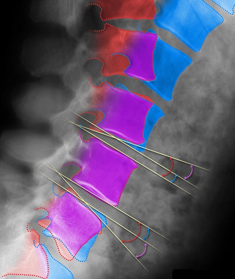
Impressive visuals for presentations


QMA® analysis visually aids in identifying changes between two X-rays, providing clinicians, scientists, and development teams with fantastic graphics for meetings and presentations.

Advanced Image Analysis

MMI’s validated, proprietary, and patented QMA® software produces highly accurate and precise measurements of change based on radiographs.
High-precision measurements of disc angle in flexion and extension using MMI’s proprietary QMA® analysis technology

What Is It?

Quantitative Motion Analysis (QMA®) is a semi-automated software algorithm used to perform image registration and accurate measurements between different x-rays of the same subject. It uses computer-assisted, image overlay methods to track the changes in position of objects (e.g. bony anatomy, device components) over time. QMA® has been shown to substantially improve accuracy and reproducibility compared to manual techniques (1,2).

How Does It Work?

QMA® uses computer-assisted, pattern-matching methods to track the objects in the imaging field of view. For example, a bony pattern or contour is identified in one image (e.g. at PreOp) and then digitally superimposed on the next image (e.g. at Follow-Up). This technique is conceptually similar to the process of overlaying two x-rays so that a specific bone is registered between the images. If landmarks are selected at PreOp, the new position of these landmarks can be calculated at Follow-Up based on the registration. The technique minimize differences in size, shape, and radiographic texture between the corresponding bony patterns on different images, and can be applied to an unlimited number of images for a given subject.

The benefit of using QMA® is that measurement landmarks are automatically propagated across all analyzed images. This avoids the reproducibility errors associated with picking landmarks on multiple images in serial radiographs and assures that the relative position of bony landmarks remains constant between images. In addition, based on MMI’s analysis techniques, certain measurements have been shown to be invariant to X-ray beam parallax effects (3).

Where’s the Evidence?

The QMA® software platform has been utilized in many trials of FDA-approved implants, including several “firsts”. Proven to increase observer reliability for common visual assessments (4), QMA® provides a proprietary visualization technique called Feature Stabilization™. Throughout the clinical trial landscape, QMA® has been cited in over 200 peer-reviewed publications and hundreds of abstracts and is widely regarded as the “gold standard” for spine quantitative image analysis. In the spine, QMA® has been tested to be at least 4 times more accurate than conventional measurement techniques (1,2), and has been validated for accuracy and reproducibility in multiple, peer-reviewed scientific studies (1,2,5,6,7,8,9). In the knee, QMA® is one of the few joint space narrowing measurement techniques that has been proven to be both reliable and accurate (10).

The QMA® technology can also be leveraged to visualize changes to any bony anatomy, device component, or other object in the field of view that conforms to rigid body assumptions. The software has additionally been used to compile authoritative databases and reference data on kinematics.

Disclaimer: QMA® is designed for investigational use only and is not for use in clinical diagnosis or patient management.

HOW IT CAN WORK FOR YOU

Applications of QMA®

Intervertebral motion measurements
(e.g. rotation, translation, center of rotation,
range of motion)
Vertebral body morphology (e.g. height, width, endplate length, augmentation)
Joint alignment (e.g. HKA angle, pelvic tilt, sagittal balance)
Joint space characterization (e.g. JSW/JSN, disc height, disc angle)
Device position (e.g. migration, subsidence, alignment angles)
Support for radiologist visual assessments (e.g. development of HO, progression of radiolucencies)

Increase in disc height between PreOp and follow-up visit after total disc replacement. C6 is held in the same position between X-rays.

Correction of scoliosis deformity after surgery. The sacrum is held in the same position between images.

Quantifiable increase in vertebral body height following vetebroplasty.

Flexion and extension X-rays after total disc replacement, showing range of motion and center of rotation (pink dot) at L4-L5. L5 is held in the same position between images.

Change in knee alignment after high tibial osteotomy. The tibia is held in the same position between PreOp and follow-up X-rays.

Some of Our Experts

Timothy Mosher, MD

Vice Chair of Radiology Research and Chief of MSK Radiology at Penn State University College of Medicine

Dr. Mosher has over 15 years’ clinical experience evaluating musculoskeletal imaging, and is a recognized expert in quantitative techniques for bone and soft tissue analysis, including cartilage relaxometry. He has published 50+ peer-reviewed papers and book chapters on musculoskeletal imaging, with an emphasis on MRI, and currently serves as the Associate Editor for Radiology, Osteoarthritis and Cartilage, Magnetic Resonance in Medicine, and is a frequent Instructional Course Lecturer for the American Academy of Orthopedic Surgeons (AAOS). Dr. Mosher is also a Member of the OARSI Working Group making recommendations to FDA on imaging techniques for OA trials.

William Morrison, MD

Professor of Radiology and Director of Musculoskeletal Radiology at Thomas Jefferson University Hospital

Dr. Morrison is an internationally recognized authority in musculoskeletal imaging with over 30 years’ experience. He has co-authored textbooks including Problem Solving in Musculoskeletal Radiology and Imaging of the Musculoskeletal System and published almost 30 book chapters and hundreds of peer-reviewed articles. Dr. Morrison has served on the editorial board of American Journal of Roentgeneology, Radiology, and Skeletal Radiology, on the Expert Panel on Musculoskeletal Radiology for the American College of Radiology (ACR), and the past president of the Society of Skeletal Radiology (SSR).

Let MMI provide insights into your clinical study imaging.

Have questions? We’ll connect you immediately to one of our scientific managers and imaging experts. Your time is precious, and we want to make the most out of it.

Contact icon
error: