Mark E. Schweitzer, MD
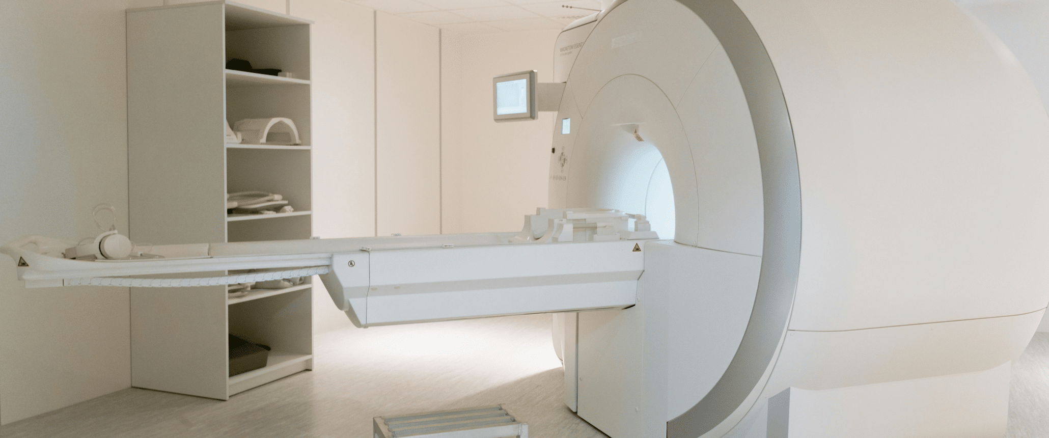
The first human MRI scanners in the 1980s operated at low magnetic field strengths below 1 Tesla (1T), which had some image quality issues primarily due to the limited signal-to-noise. By the early 1990s, 1.5T became standard across healthcare centers. Since then, functional sequences requiring even higher spatial and temporal resolution have been developed, and thus, many imaging centers have progressed to 3T imaging. Even higher field strengths (5T and more recently, 7T) have been piloted, though these have not taken off, often due to space, cooling, and weight issues, in addition to expense and sensitivity in implementing day-to-day operations.
Concurrent with the transition to higher field strengths, more powerful gradients have come into play, extending the maximum spatial resolution of the MRI scanner even further. About 20 years ago, academic researchers proposed incorporating these more powerful gradients on lower field scanners but received pushback partially related to marketing and demand concerns.
Worldwide trends in the last five (5) years have put global warming in sharp focus in radiology. Radiologists and MRI system manufacturers have become increasingly concerned with both power and volatile gas usage in higher field strength MRI systems. These resource needs notably limit the wide-spread adoption of high-field MRI usage. Lower field scanners typically use permanent magnets without the need for cryogens and have lower energy, shielding, and cooling requirements.
More recently, many thought leaders have circled back to low-field strength MRI scanners. With recent AI advances coupled with the use of stronger gradients and advancements in MRI coil technology, it has become possible to achieve high-field-comparable image quality using lower field strength. Therefore, major vendors have begun to shift towards low-field magnets. In this permutation with AI assistance, functional sequences such as diffusion imaging can now be offered at lower cost.
Studies have found that with these optimizations at lower field strengths, both objective and subjective assessments demonstrate non-inferiority. As trends continue, lower field scanners will likely diffuse rapidly across the healthcare ecosystem so that pivotal clinical trial protocols can be used across a wider range of geographies and practice settings. This diffusion will allow MRI to be performed in non-traditional environments, including rural areas, developing nations, small physician offices, and even sports stadiums with mobile MRI units. The best applications are to the knee, wrist, shoulder, elbow, and perhaps the ankle.
While research into higher field strengths will undoubtedly continue for niche areas such as pre-clinical imaging, there is a slow but steady shift back to low-field MRI.
Islam, K.T., Zhong, S., Zakavi, P. et al. Improving portable low-field MRI image quality through image-to-image translation using paired low- and high-field images. Sci Rep 13, 21183 (2023). https://doi.org/10.1038/s41598-023-48438-1
Hennig J. (2023). An evolution of low-field strength MRI. Magma (New York, N.Y.), 36(3), 335–346. https://doi.org/10.1007/s10334-023-01104-z
Liu, H., Liu, J., Li, J., Pan, J. S., & Yu, X. (2021). DL-MRI: A Unified Framework of Deep Learning-Based MRI Super Resolution. Journal of healthcare engineering, 2021, 5594649. https://doi.org/10.1155/2021/5594649
Dr. Mark Schweitzer is Vice President of Health Affairs at Wayne State University and Editor-in-Chief of the Journal of Magnetic Resonance Imaging (JMRI). He is a key opinion leader in the field of medical imaging and one of MMI’s scientific advisors. He has published 400+ peer-reviewed articles and 60+ book chapters.
Medical Metrics, Inc. is an ISO 9001:2015-certified provider of independent imaging core lab services and a leading core lab for clinical trials and related consulting. We assist sponsors with designing, implementing, and executing the imaging strategy of their global trials through our responsive trial management team, robust operating infrastructure, and world-class imaging expertise. Learn more about our Therapeutic Area expertise and our Services.


