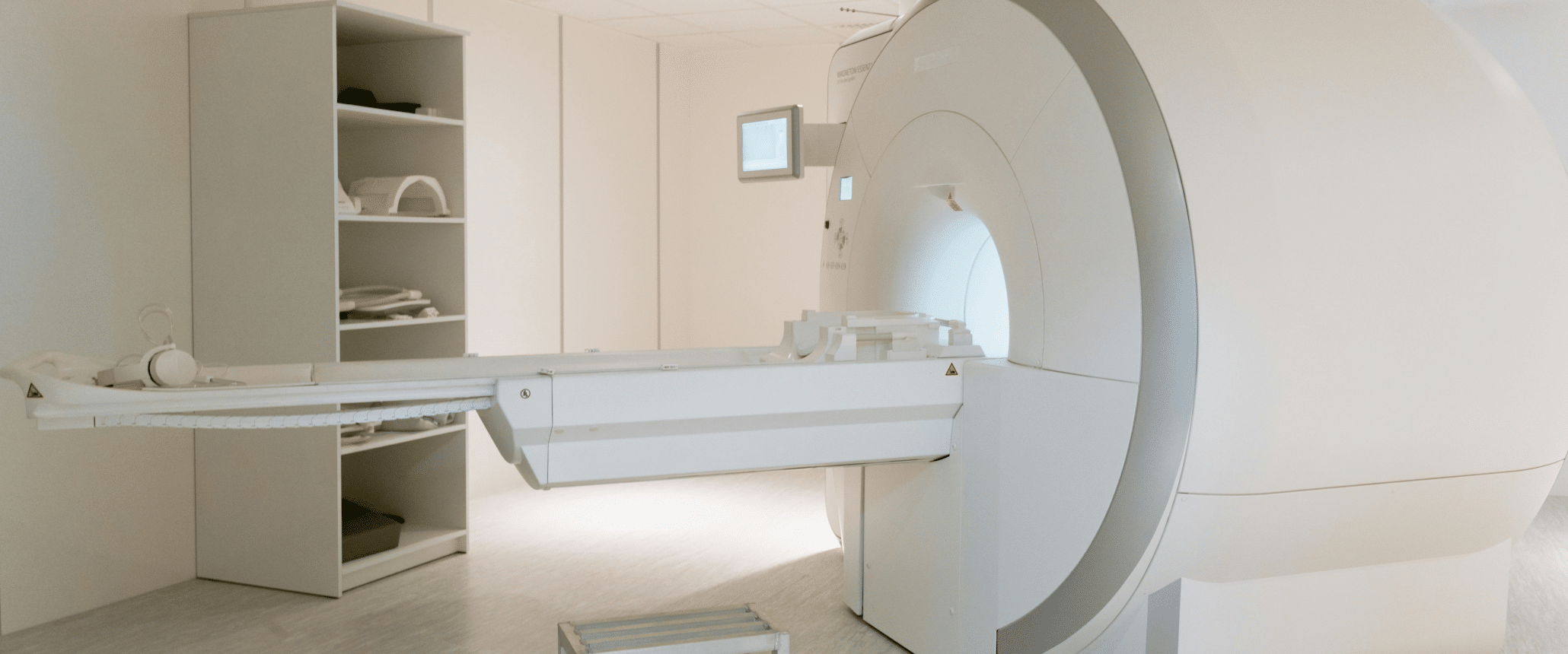
Imaging plays a key role in patient selection for GAE treatment.
Key Highlights:
- Mild OA Patients May Respond Better to GAE: A recent study by Gill et al. found that patients with KL Grade 2 (mild OA) showed significantly greater improvements in pain and function compared to those with KL Grades 3–4 (moderate/severe OA) [1].
- MRI Biomarkers Matter: Baseline MRI features such as full-thickness cartilage defects, effusion synovitis, bone marrow lesions, and osteophytes correlate with reduced pain relief post-GAE [2].
- Trials Require Precision: With median musculoskeletal drug trial costs exceeding $41,000 per subject [3], enrolling the right patients is critical. Imaging-based selection criteria can reduce trial failure risk and improve data clarity.
What is GAE?
Genicular artery embolization (GAE) trials have gained noticeable traction in recent years, particularly for treating knee osteoarthritis (OA) pain in patients who don’t respond to conservative treatments or aren’t candidates for surgery. The procedure’s minimally invasive nature and promising early results have sparked interest, but it’s still considered an emerging therapy with a growing but not yet widespread research footprint.
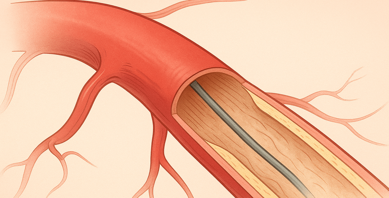
- GAE is a minimally invasive intervention for mild-to-moderate knee OA patients refractory to conservative treatments, and not considered candidates for total knee replacement. A catheter is inserted into the genicular artery (via the femoral artery) to deliver microspheres, selectively occluding neovessels thought to drive synovial inflammation and pain.
- First explored for knee hemarthrosis, GAE’s use for OA pain expanded after Dr. Yuji Okuno’s 2014 study demonstrated rapid pain relief in OA patients [4].
Research and Trials: Key Findings
- KL Grade and Patient Reported Outcome (PRO): A recent study conducted by Gill et al., found patients with a baseline Kellgren-Lawrence (KL) Grade of 2 (mild radiographic OA) demonstrated greater improvements in pain, function, and quality of life after GAE when compared with patients with baseline KL Grades of 3 (moderate/severe radiographic OA) [1]. There were no KL Grade 4 (severe radiographic OA) responders, thus indicating poor PROs for patients with severe knee OA.
- MRI’s Role in Biomarker Identification: Zadelhoff et al. identified full-thickness cartilage defects as the strongest MRI-based predictor of reduced pain relief post-GAE. Other markers like effusion synovitis and bone marrow lesions also correlated with poor outcomes [2].
- Need for Controlled Trials: Experts agree, current evidence lacks high-quality, randomized, sham-controlled trials with long-term follow-up. Such studies are critical to confirm GAE’s efficacy and refine patient selection criteria [5]
Why Imaging Matters in GAE Trials
- Imaging Biomarkers Guide Eligibility: MRI and X-ray data must be rigorously analyzed to exclude patients with signs of advanced OA (e.g., KL Grade 4, severe cartilage defects) that may limit treatment benefit.
- Cost Efficiency: Poor patient selection risks trial failure and wasted resources. Imaging-driven criteria ensure only the most likely responsive populations are enrolled, reducing attrition and improving data validity.
How MMI Can Help You Succeed
- Protocol-Driven Inclusion/Exclusion Criteria: Leverage our experience in 200+ OA trials to design imaging-based criteria that align with biomarker evidence (e.g., KL grades, MRI features).
- Independent Image Analysis: Ensure unbiased assessment of X-ray and MRI data to confirm patient eligibility and adherence to trial protocols.
- Quality Control: Our standardized workflows minimize variability, ensuring your data meets regulatory and publication standards.
- Data-Driven Decisions: Access actionable insights to refine trial design, optimize enrollment, and reduce costs.
Contact our imaging experts today to learn how MMI’s core lab services can strengthen your trial’s rigor and success.
Medical Metrics, Inc. is an ISO 9001:2015-certified provider of independent imaging core lab services and a leading core lab for orthopedics clinical trials and related consulting. We assist sponsors with designing, implementing, and executing the imaging strategy of their global trials through our responsive trial management team, robust operating infrastructure, and world-class imaging expertise.
[1] Gill SD, Hely R, Hely A, Harrison B, Page RS, Landers S. Outcomes after Genicular Artery Embolization Vary According to the Radiographic Severity of Osteoarthritis: Results from a Prospective Single-Center Study. J Vasc Interv Radiol. 2023 Oct;34(10):1734-1739.
[2] van Zadelhoff TA, Okuno Y, Bos PK, Bierma-Zeinstra SMA, Krestin GP, Moelker A, Oei EHG. Association between Baseline Osteoarthritic Features on MR Imaging and Clinical Outcome after Genicular Artery Embolization for Knee Osteoarthritis. J Vasc Interv Radiol. 2021 Apr;32(4):497-503.
[3] Moore TJ, Heyward J, Anderson G, Alexander GC. Variation in the estimated costs of pivotal clinical benefit trials supporting the US approval of new therapeutic agents, 2015-2017: a cross-sectional study. BMJ Open. 2020 Jun 11;10(6):e038863.
[4] Okuno Y, Korchi AM, Shinjo T, Kato S. Transcatheter arterial embolization as a treatment for medial knee pain in patients with mild to moderate osteoarthritis. Cardiovasc Intervent Radiol. 2015 Apr;38(2):336-43.
[5] Little MW, Motta-Leal-Filho JM, Okuno Y. Panel Discussion: Genicular Artery Embolization: Building Evidence and Practice. Endovascular Today. 2024 Feb;Vol.23,No.2.
Latest Scientific Resources & Publications
MMI CSO, Dr. John Hipp Selected as Guest Editor of Bioengineering Special Issue! - Submissions Due Nov 30
Why You Need an Imaging Core Lab: 4 Ways a Commercial ICL Transforms Your Trial


