MMI Co-Founder, John Hipp, PhD. to be Guest Editor for Special Issue of Bioengineering! Submit by May 30!
Learn MorePress Release!: Driving Excellence in Cardiovascular Trials: MMI and Healthcare Inroads Deepen Collaboration
Read HereThe MMI Advantage
100+ unique orthopedic treatments evaluated
MMI’s breadth of experience ranges from soft tissue repair and regeneration to hemi and total joint replacements.
Validated and accurate JSW/ JSN measurement technique
Our joint space narrowing measurement technique has been validated for both accuracy and reproducibility using our patented QMA® technology.
Experienced with all major joints in the body
We have evaluated all of the major articular joints in the body, including in the hands and feet.
Experience and Expertise
MMI has over fifteen years of experience in providing core lab support for more than 150 orthopedic device and biopharmaceutical companies internationally, including 14 of the top 15 global market leaders.
Modality Expertise
X-ray
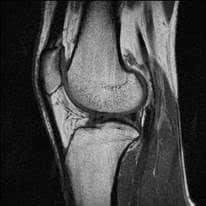
MRI
CT
Ultrasound
At MMI, we have evaluated well over 100 unique treatments for musculoskeletal disorders of the hip, knee, ankle, wrist, shoulder, elbow, peripheral joints, and long bones involving osteoarthritis, rheumatoid arthritis, trauma, and sports medicine. Broadly, our musculoskeletal treatment experience includes but is not limited to drugs, stem cells, biologics, viscosupplements, marrow stimulation, tissue grafting, ablation, bone fillers, fixation devices, bioresorbable scaffolds, biphasic implants, biomimetic devices, resurfacing, and partial and total arthroplasty.
WHAT WE OFFER
Analysis Capabilities
Tissue structure, morphometry, and hydration (e.g. thickness, volume, lesion depth, T2 and T1ρ relaxation mapping)
Soft tissue repair & regeneration (cartilage, meniscus, tendon, ligament, muscle)
Joint space width and narrowing
Joint stability and alignment
Whole joint health semi-quantitative scoring systems
Implant stability, fixation, and alignment
Bone-implant and cement-implant interface remodeling
Structural bony anatomy (e.g. osteophytes, cysts, HO)
Fracture reduction, stabilization, and healing
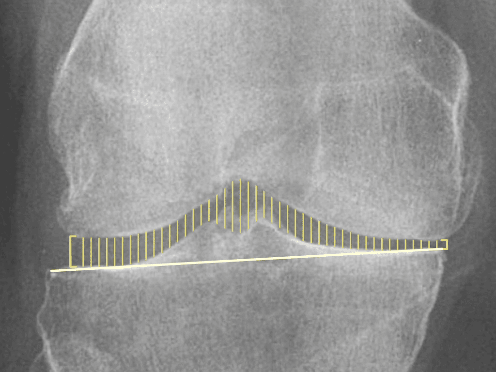
Radiographic joint space width using the mid-coronal plane measurement method
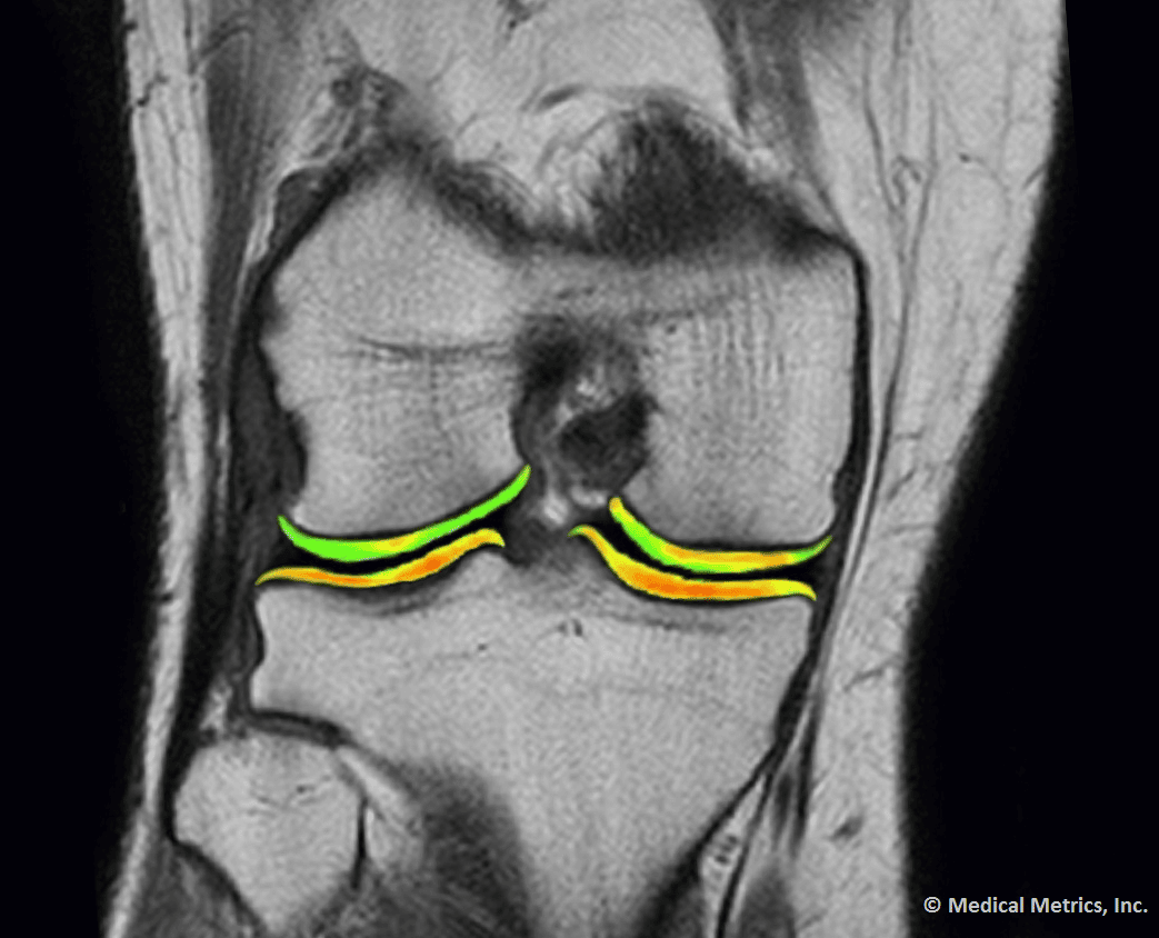
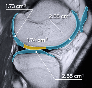
MR relaxation mapping of knee cartilage
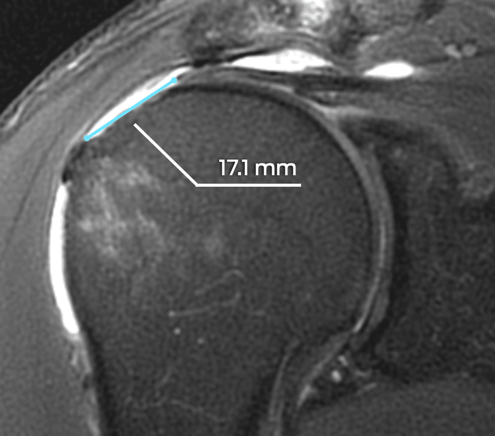
Preoperative rotator cuff tear length on sagittal MR
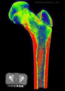
Femoral bone density analysis from CT
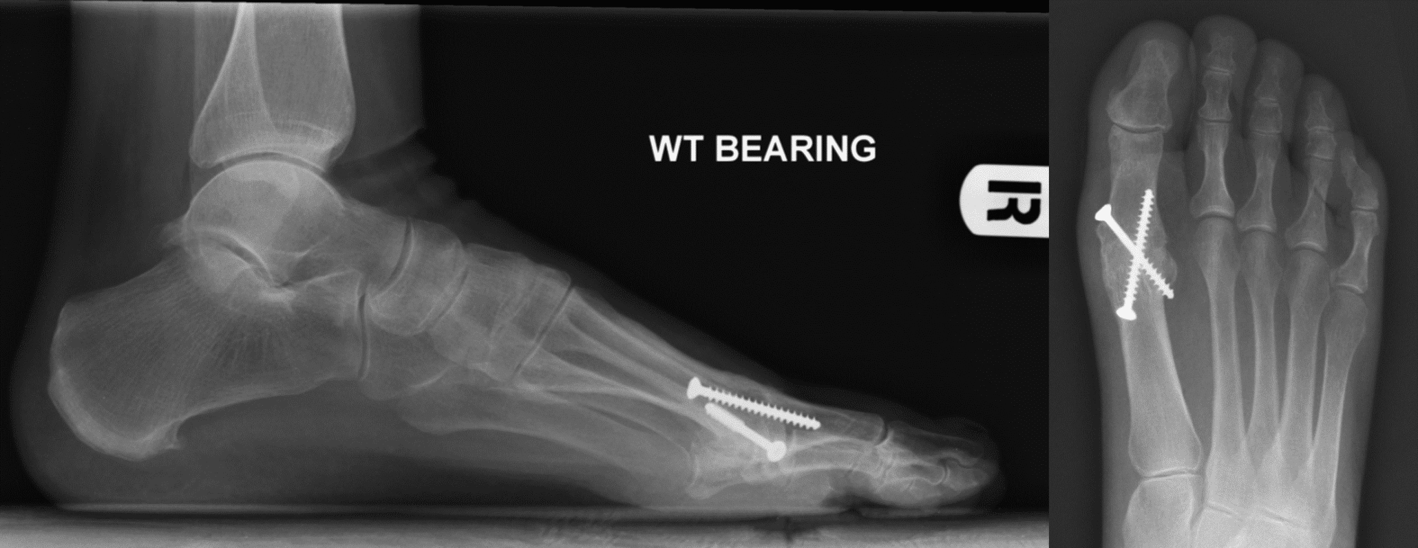
Successful fusion of the MTP at 24 months
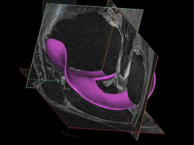
3D surface of cartilage created from an MRI
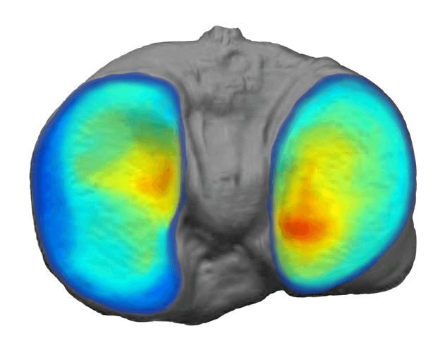
Tibial cartilage thickness mapping
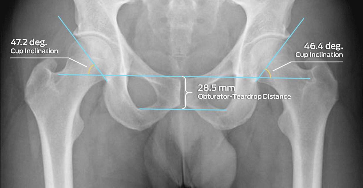
Acetabular cup inclination and obturator-tear drop distance measurements to assess coverage and pelvic positioning, respectively
Some of Our Experts
Timothy Mosher, MD
Kenneth L. Chair, Department of Radiology and Distinguished Professor of Radiology and Orthopaedic Surgery at Penn State University
Dr. Mosher is a clinical musculoskeletal radiologist with a research interest in osteoarthritis. He is a fellow of the International Society for Magnetic Resonance in Medicine (ISMRM) and has served on numerous NIH scientific review groups, the Scientific Advisory Board for the Canadian Arthritis Network, the Canadian Foundation for Innovation, and as an industry consultant in the areas of imaging applications to musculoskeletal projects. Dr. Mosher has published over 80 books and manuscripts with the majority in the area of musculoskeletal imaging, quantitative cartilage imaging, and MRI technical development.
William Morrison, MD FACR
Professor of Radiology and Director of Musculoskeletal Radiology at Thomas Jefferson University Hospital
Dr. Morrison is an internationally recognized authority in musculoskeletal imaging with over 30 years’ experience. He has co-authored or edited 13 textbooks including Problem Solving in Musculoskeletal Radiology and Imaging of the Musculoskeletal System, and published 100 book chapters and over 150 peer-reviewed articles. Dr. Morrison has served on the editorial board of the journals Radiology, Skeletal Radiology, and Seminars in Musculoskeletal Radiology. He has served on Expert Panels for the American College of Radiology (ACR), the American Board of Radiology (ABR), and the American Diabetes Association (ADA). He is also a past president of the Society of Skeletal Radiology (SSR).
Relevant Scientific Resources
Key Publications Involving MMI
Let MMI provide insights into your clinical study imaging.
Have questions? We’ll connect you immediately to one of our scientific managers and imaging experts. Your time is precious, and we want to make the most out of it.
